 | ||||
How to Prepare a Wet Mount Slide of
Eukaryotic Cells
In order to view small objects with a compound microscope, the object, or specimen must be properly prepared, mounted on a slide, and sometimes stained to increase the contrast between the object and background.
Article Summary: Preparing a wet mount of a specimen is the technique typically used to view plant and animal cells using a microscope. This page provides step by step instructions on slide preparation as well as videos at the bottom of page.
How to Prepare a Wet Mount Slide
Page last updated: 1/2016
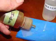
Step Five - If the specimen is transparent, such as onion skin or check cells, stain should be added to increase contrast. A drop of iodine or methylene blue is commonly used. Do not use stain if viewing photosynthetic cells (which already appear green due to chlorophyll), or living organisms, such as protozoans in pond water (stains will kill them).
SPO VIRTUAL CLASSROOMS
 | ||||||
How to Prepare a Wet Mount Specimen
Step One - Obtain a clean microscope slide.
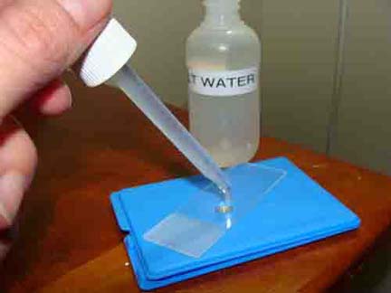
Step Two - Place a drop of liquid on the slide. This is the “wet” part of the wet mount. The liquid used depends on the type of cell being viewed:
- If examining a plant cell, tap water can be used.
- If examining an animal cell, physiological saline (or contact lens solution) must be used, because if plain water is used, the cell will explode from osmotic pressure. Unlike plant cells and bacteria, animal cells have no cell wall to structurally support them.
Step Three - Obtain the specimen to be used. Some introductory biology classics for viewing include:
- Skin of an onion bulb: In order to view the cells, a very thin layer of skin must be obtained. Take a single layer of onion and bend it towards the shiny side. After it snaps, pull gently, and a transparent layer of skin, similar to Scotch tape, will appear.
- Elodea leaf: Elodea leaves are two cell layers thick. The cells in one layer are smaller than the cells in the other, so elodea leaves can be used to better understand a microscope's depth of field.
- Cheek cells: Human epithelial cells can be obtained by gently rubbing a toothpick on the inside of the mouth, and then swirling the toothpick in the physiological saline on the slide.
- Pond water: Obtaining some water from a pond makes wet mount preparation a breeze, since the water and the specimens are both included.
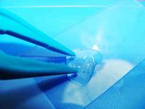
Step Four - Place the specimen into the liquid on the slide.
SPO VIDEO Tutorial on Compound Light Microscope Parts & Operation
2.
4.
5.
6.
Wet mount technique is used for preparing eukaryotic cells, such as the cells of plants and animals for the microscope. In order to view bacteria (prokayotes), which are much smaller than plant and animal cells, a different specimen preparation technique is used called a bacterial smear.
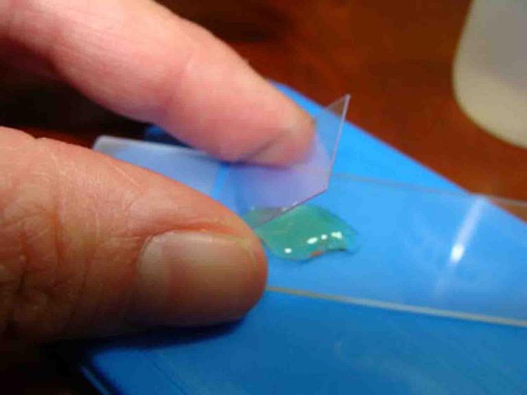
Step Six - Place a cover slip over the specimen. This will sandwich the specimen between the slide and the cover slip. To avoid trapping air bubbles, set one edge of the cover slip on the slide, and then let the rest of the cover slip drop. As it drops from one side to the other, air will be pushed out, and this will reduce the number of bubbles in the wet mount.
You have free access to used in a college-level introductory Cell Biology Course. The Virtual Cell Biology Classroom provides a wide range of free educational resources including Power Point Lectures, Study Guides, Review Questions and Practice Test Questions.
