 | ||||
How to Prepare a Wet Mount Slide - P2
Page last updated: 4/2015
Viewing a Prepared Microscope Slide
The specimen is now ready to be viewed through the microscope. Once the slide is positioned on the microscope stage, start at a low power (4x or 10x) objective, and work up to the level of magnification required to get a detailed view of the specimen.
Remember to adjust the level of light to increase the contrast and make your specimen easier to view.
SCIENCE PHOTOS
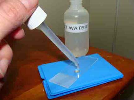
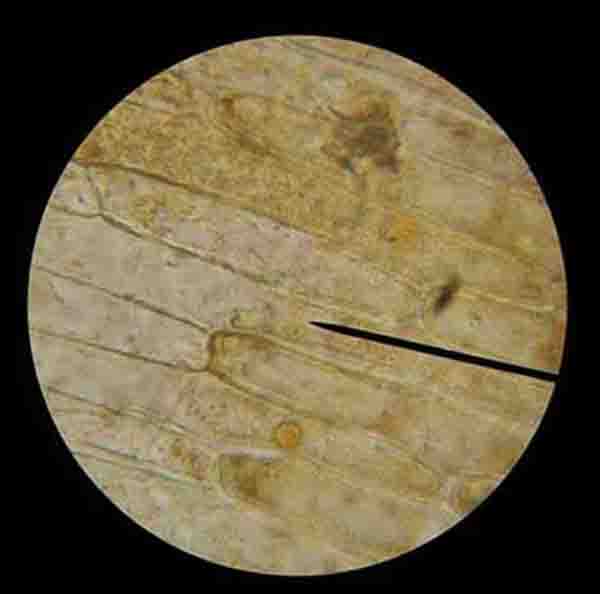
ABOVE: Photo of wet mount of onion skin specimen being prepared. BELOW: Onion cells viewed through microscope @ 400xTM.
SPO VIRTUAL CLASSROOMS
SPO VIDEO: How to Make a Wet Mount of Stained Epithelial Cheek Cells
SPO VIDEO: How to Make a Wet Mount Slide of Stained Onion Skin Cells
SPO VIDEO: How to Make a Wet Mount Slide of
Elodea Plant Cells
 | ||||
PAGE 2 < Back to Page 1
Sources & Resources
- "How to Use a Compound Microscope". Tips on how to find and focus on microscopic specimens.
- Microscopy Laboratory Exercise Main Page from the Virtual Microbiology Classroom.
- Eukaryotic Cell Structure Lecture Main Page from the Virtual Cell Biology Classroom.
- Virtual Microscope from the University of Delaware.
VIDEOS OF WET MOUNT TECHNIQUE
FOR VIEWING CELLS
Page last updated: 1/2016
The Virtual Cell Biology Classroom provides a wide range of free educational resources including Power Point Lectures, Study Guides, Review Questions and Practice Test Questions.


